Protein AI field guideArticle 3 of 7
Choose the Site Before the Binder
Trastuzumab and pertuzumab engage different surfaces of HER2. Their measured structures show why the location of binding belongs in the design brief before affinity becomes the optimization target.
- Evidence type
- Published-study walkthrough
- Published
- August 2026
The surface is part of the therapeutic hypothesis
Two binders can recognize the same receptor and do different jobs. One may block a partner; another may leave that interaction intact. A positive binding assay tells a team that a molecule engages its target. It does not, by itself, tell them whether it engages the useful surface.
At Crane, protein engineers turn the intended mechanism into a spatial brief: the receptor feature to block or engage, the surfaces to avoid, the orientation a payload may need, and the measurements that would prove the hypothesis.
One receptor, two measured intervention sites
Select an epitope to see how the biological hypothesis changes.
Loading measured HER2 complex
Drag to rotate · scroll to zoom
Rose: domain IV · orange: domain II
Epitope map
One target supports different hypotheses
The same measured HER2 ectodomain carries two spatially distinct antibody epitopes. Choosing between them changes which receptor event a new binder could influence.
Evidence boundary
Co-occupancy establishes compatible geometry. It does not establish combination benefit, additivity, or clinical effect.
Measured reference: PDB 8Q6J
The same receptor presents different intervention sites
The measured cryo-EM structure PDB 8Q6J captures one HER2 ectodomain with both trastuzumab and pertuzumab Fabs. It makes the strategic contrast visible on the same receptor.
Trastuzumab occupies the membrane-proximal domain IV epitope also measured in PDB 1N8Z. Pertuzumab binds near the center of domain II in PDB 1S78, beside a receptor dimerization surface.
The pertuzumab structure and mutagenesis study supports a steric model for interfering with receptor dimerization. The structure does not establish that any new binder at that site will reproduce the same affinity, signaling effect, or cellular response.
| Example | Measured binding site | Design implication |
|---|---|---|
| Trastuzumab | A membrane-proximal HER2 domain IV epitope. | Site and orientation must be assessed in the context of the intended mechanism and format. |
| Pertuzumab | A domain II epitope beside a receptor dimerization surface. | The published structural and mutational evidence supports a steric mechanism. |
| A proposed binder | An interface predicted on the chosen surface. | Competition and functional measurements must test whether engagement produces the intended effect. |
HER2 domain IV is closest to the membrane; domain II sits farther along the ectodomain. Rose marks the trastuzumab site, orange the pertuzumab site.
Domain order only, not to scale. The membrane is a contextual cue, not measured coordinates or an orientation fitted to the rotatable structure.
Conceptual illustration
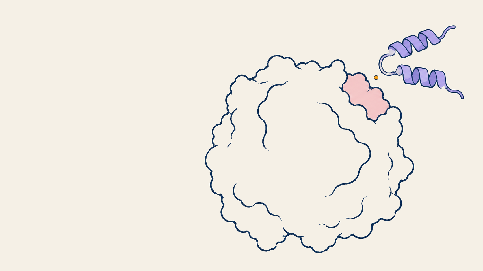
A binding site needs a biological reason
A generic target and a proposed binder illustrate the choice of surface. The biological question determines which encounter is worth testing.
Site selection comes before affinity optimization
Once the intended receptor event is clear, an epitope can become a design constraint. Hotspots can define where contacts should form. Negative constraints can keep a candidate away from glycans, conserved family surfaces, or geometry that would obstruct the intended format.
A published HER2 domain IV binder program used iterative computational design and experimental feedback to refine a site-specific LRR binder. That loop matters. It is not a one-shot demonstration that choosing a patch and running a model produces a development candidate.
The deliverable is a spatial brief that explains why this surface is worth pursuing. It names the accessibility and orientation constraints and the result that would make the team reject the proposed mechanism. A predicted interface then carries a testable argument into the synthesis decision.
How an epitope becomes an experimental plan
01 · Define
Name the receptor event
Dimerization, internalization, recruitment, steric blockade, or payload positioning each imply a different spatial brief.
02 · Constrain
Mark required and forbidden surfaces
Encode hotspots, orientation, accessibility, glycan context, and selectivity risks before generating candidates.
03 · Challenge
Test the interface as an ensemble
Use orthogonal models and structural checks to find collisions, unstable contacts, and alternative poses before synthesis.
04 · Prove
Connect binding to biology
Measure binding, competition, signaling, trafficking, and cellular response in the context the mechanism requires.
Related Crane work: a computational PD-L1 study
Our PD-L1 cyclic-peptide study shows how we evaluated a different interface problem computationally. It reports design and structural assessment, not measured binding or a validated therapeutic outcome. It is an example of Crane's execution alongside the published HER2 evidence discussed here.
Which surface would change the biology?
Bring your target and intended mechanism. We can turn them into an epitope brief, assess accessibility and competing poses, and define the evidence a candidate needs before your team commits to synthesis.
Part 3 of 7
Protein AI field guide
Protein AI in practice: a seven-part field guide
Seven connected decisions, illustrated by different molecular systems and published studies. Frame the problem and choose a route, specify the physical constraints, then decide what evidence earns the next experiment.
Start with the decision in front of you. After choosing a state, follow the binding-site or catalytic-chemistry example that fits your application. Article numbers follow publication order.
- 01
Frame the problem
Available now
Map the Molecular System
Before models run, map the constructs, partners, ligands, stoichiometry, and interfaces that carry the biology.
- 05
Available now
One Target, Three Development Strategies
Proteins, peptides, nucleic acids, and small molecules offer different routes to the same goal.
- 02
Specify the design
Available now
The Target Changes Shape
Compare measured target states before choosing a structural reference.
- 03
Current story3 of 7
Choose the Site Before the Binder
For binding programs: connect the chosen surface to the intended biological effect.
- 04
Available now
Design the Chemistry Before the Scaffold
For enzyme programs: distinguish a catalytic geometry from measured activity.
- 06
Decide what advances
Available now
When a Binder Earns the Next Experiment
Separate measured binding, solution behavior, and structural agreement from open development questions.
- 07
Learn from measurement
Available now
The Next Round Starts with the Assay
Build, test, learn, and feed experimental results into the next design round.