Protein AI field guideArticle 1 of 7
Map the Molecular System
A Crane reconstruction recovered the known BRD4-MZ1-pVHL interface with two system definitions. Adding the surrounding partners did not consistently improve the scores. The comparison shows how to challenge an assembly choice before committing to experiments.
- Evidence type
- Crane reconstruction
- Published
- July 2026
Why the system map comes first
A degrader can bind both of its partners and still fail to arrange them productively. For a discovery team, that is a costly ambiguity: should the next experiment test the compound, the protein constructs, or the assumed mechanism? A map of the complete molecular system helps separate those questions before the assay queue fills up.
The 5T35 crystal structure captures one such system. BRD4 binds the PROTAC MZ1, MZ1 binds VHL, and the two proteins create contacts that do not exist in either binary complex.
We compared a minimal assembly with a fuller one to ask a specific question: would including the surrounding partners change recovery of the interface? That comparison tests a modeling choice; it does not establish whether a new degrader will work.
This is a known-case reconstruction. The 2017 release of 5T35 falls before Boltz-2's structural training cutoff, and the reference is not a documented holdout. It cannot establish generalization to unseen systems. The value here is a traceable comparison of two assembly definitions.
Loading molecular system
Drag to rotate · scroll to zoom
Assembly walkthrough
Start with the target
Experimental structure loaded: BRD4 is shown in isolation.
Evidence boundary
A binary view cannot tell us how an E3 ligase will approach the target.
Violet and teal show the experimental 5T35 assembly. The pale cyan overlay shows the independently generated Boltz-2 reconstruction after sequence-aware alignment.
What the map revealed in a known PROTAC system
Both specifications recovered the known protein interface in all five samples. DockQ measures agreement with an experimental protein interface on a scale from 0 to 1, with higher values indicating closer agreement. It is a reference-based accuracy metric, not a model confidence score.
The minimal specification contained BRD4 BD2, pVHL, and MZ1. The full specification added ElonginB and ElonginC. We used the same computational protocol for both, with five samples per system.
The five minimal-system structures scored 0.903 to 0.970 DockQ for the induced BRD4-pVHL interface. The five full-system structures scored 0.922 to 0.940. Each recovered 13 to 15 of the 15 native protein contacts.
MZ1 also returned to the experimental pose. Its symmetry-aware heavy-atom RMSD was 0.6 to 1.4 Å after aligning the relevant protein pocket. For the retrospective visual, we show full-system sample 1, the highest-DockQ full-system pose. It aligns to the measured assembly at 0.892 Å across 451 matched protein C-alpha atoms.
These are ten correlated samples from one known case and a shared seed, not ten independent validation cases. Both biological specifications recovered the interface; the full system was not automatically more accurate than the reduced one.
Five samples per system, compared with the same 5T35 reference. Each dot is one recorded BRD4-pVHL DockQ score. Higher is closer to the measured interface.
Detail: 0.88 to 1.00 of the full DockQ scale (0 to 1)
One known case, with correlated samples from a shared seed. Vertical offsets separate the dots; they do not represent another measurement.
Inspect the ten sample scores
| System | Sample | DockQ |
|---|---|---|
| Minimal assembly | 0 | 0.903 |
| Minimal assembly | 1 | 0.921 |
| Minimal assembly | 2 | 0.970 |
| Minimal assembly | 3 | 0.904 |
| Minimal assembly | 4 | 0.941 |
| Full assembly | 0 | 0.936 |
| Full assembly | 1 | 0.940 |
| Full assembly | 2 | 0.934 |
| Full assembly | 3 | 0.922 |
| Full assembly | 4 | 0.925 |
| What was compared | Observed in this reconstruction | What it supports |
|---|---|---|
| Minimal assembly | Five samples; BRD4-pVHL DockQ 0.903 to 0.970. | The reduced system recovered the known interface. |
| Full assembly | Five samples; BRD4-pVHL DockQ 0.922 to 0.940. | Adding ElonginB and ElonginC retained the interface without improving every score. |
| MZ1 pose RMSD | 0.6 to 1.4 Å across both ensembles after pocket alignment. | The ligand pose was close to the measured reference. Binding and degradation still require experiments. |
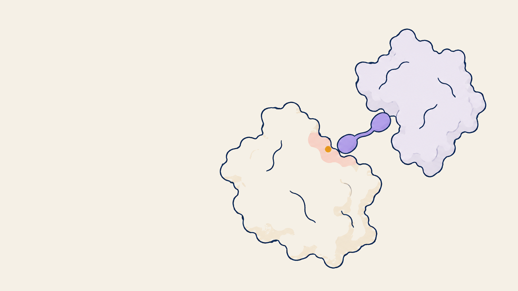
Conceptual illustration
The partners are part of the question
A system map makes molecular relationships explicit. This generic assembly sketch frames the choice of partners; the reconstruction above compares actual structures.
What this changes in the design brief
Here, adding the two partners did not change the central structural conclusion. A team can use that comparison to decide whether the extra assembly context is needed for its question, rather than assuming that a larger input is always a better one.
For a novel system without a measured reference, DockQ and reference ligand RMSD are unavailable. Confidence scores cannot substitute for them. The design brief must say which predicted contacts or assembly assumptions an experiment will test.
The map also helps choose where to intervene. Blocking a circulating protein, changing its production, and recruiting another partner lead to different programs. The next article in the reading guide examines that choice of molecular route.
How the map becomes an experimental plan
The original 5T35 study did the work a structural model cannot. The authors measured binary and ternary binding, quantified cooperativity, mutated interface residues, and connected the structural interface to degradation behavior. Read the primary experimental study.
For a new system, we would use the predicted assembly to choose interface mutations and orthogonal assays. SPR can quantify binary and ternary binding, kinetics, and cooperativity; BLI can provide an orthogonal binding readout. Mutagenesis can test whether the proposed contacts matter. Cellular degradation can test whether the assembled geometry produces the intended phenotype.
Evidence ledger
Measured
5T35 is a 2.7 Å experimental crystal structure. The published study measured ternary binding, cooperativity, mutational effects, and degradation.
Computed
On the pre-cutoff 5T35 reference, two controlled five-sample runs generated ten computational samples. Each recovered a high-quality BRD4-pVHL interface and the experimental MZ1 pose.
Capability context
Boltz-2's published methods describe a structural training cutoff after 5T35's 2017 release, so this is a known-case reconstruction rather than a holdout benchmark. The value is the worked example: a traceable system definition, an explicit comparison with measurement, and a clear account of what to test next. This case does not establish generalization to unseen molecular systems.
Methods and sample data
Both runs used Boltz 2.2.1, stereochemical SMILES for MZ1, MMseqs2 MSAs, seed 42, 10 recycling steps, 200 diffusion steps, and five samples. The 5T35 coordinates were used for evaluation after inference, not supplied as inputs. Settings were evaluated together, so this comparison cannot attribute the outcome to any one setting. Read the sample-level data and alignment methods.
Which assembly should your team test?
Bring your target, known partners, and the decision holding up the next experiment. We can scope a system map, a comparison of plausible assemblies, and the evidence needed to choose between them.
Part 1 of 7
Protein AI field guide
Protein AI in practice: a seven-part field guide
Seven connected decisions, illustrated by different molecular systems and published studies. Frame the problem and choose a route, specify the physical constraints, then decide what evidence earns the next experiment.
Start with the decision in front of you. After choosing a state, follow the binding-site or catalytic-chemistry example that fits your application. Article numbers follow publication order.
- 01
Frame the problem
Current story1 of 7
Map the Molecular System
Before models run, map the constructs, partners, ligands, stoichiometry, and interfaces that carry the biology.
- 05
Available now
One Target, Three Development Strategies
Proteins, peptides, nucleic acids, and small molecules offer different routes to the same goal.
- 02
Specify the design
Available now
The Target Changes Shape
Compare measured target states before choosing a structural reference.
- 03
Available now
Choose the Site Before the Binder
For binding programs: connect the chosen surface to the intended biological effect.
- 04
Available now
Design the Chemistry Before the Scaffold
For enzyme programs: distinguish a catalytic geometry from measured activity.
- 06
Decide what advances
Available now
When a Binder Earns the Next Experiment
Separate measured binding, solution behavior, and structural agreement from open development questions.
- 07
Learn from measurement
Available now
The Next Round Starts with the Assay
Build, test, learn, and feed experimental results into the next design round.