De Novo Nanobody Design for a Bispecific BBB Shuttle: 32 Variants to Synthesis
A computationally designed bispecific VHH panel: a TfR1-binding arm validated against the full receptor across two independent structure predictors, plus an alpha-synuclein fibril-binding arm. 100 candidates funneled to 32 synthesis-ready variants. No wet-lab validation yet.
- Evidence type
- Use Case
- Published
- April 2026
What was designed, how, and what remains unvalidated
Task
De novo design of bispecific VHH: TfR1-binding arm (apical-domain transcytosis epitope) + aSyn fibril-binding arm (NAC core epitope)
Inputs
TfR1 crystal structure (PDB 1SUV), aSyn fibril cryo-EM structures (NAC core residues 68-79), common VHH framework scaffold
Design method
IgGM (diffusion-based CDR co-design), 50 candidates per target, 100 total. Zero borrowed CDR sequences.
Validation
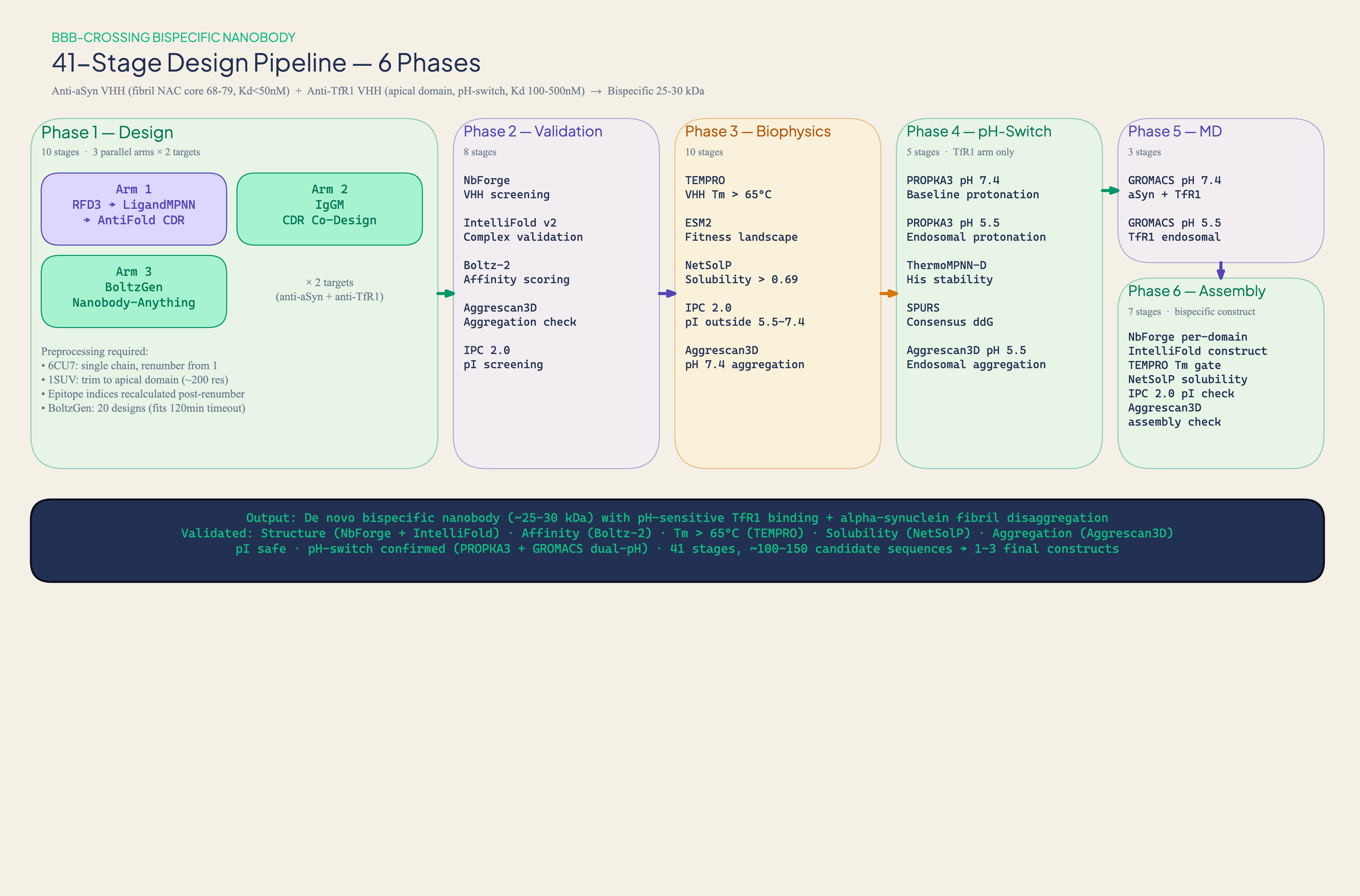
11 tools across 8 quality gates: NbForge, TEMPRO, IPC 2.0, IntelliFold v2, Boltz-2, Aggrescan3D, PROPKA3, ThermoMPNN-D, plus a full-receptor steric check against the intact 1SUV assembly
Output
32 synthesis-ready variants (2 TfR1 binders validated across both predictors, 30 aSyn top binders)
Status
Computational only. No wet-lab validation yet. SPR, ThT, DSF, SEC-MALS pending.
Purpose
This is a capability demonstration, not a therapeutic development program. The goal is to show the depth and rigor of the computational workflows we deliver as services, not to advance these specific candidates into the clinic.
The challenge
Parkinson's disease kills dopaminergic neurons. Alpha-synuclein fibrils, toxic protein aggregates, spread through the brain like an infection, seeding aggregation as they go. The therapeutic logic is straightforward: clear the fibrils, slow the disease. The engineering challenge is anything but.
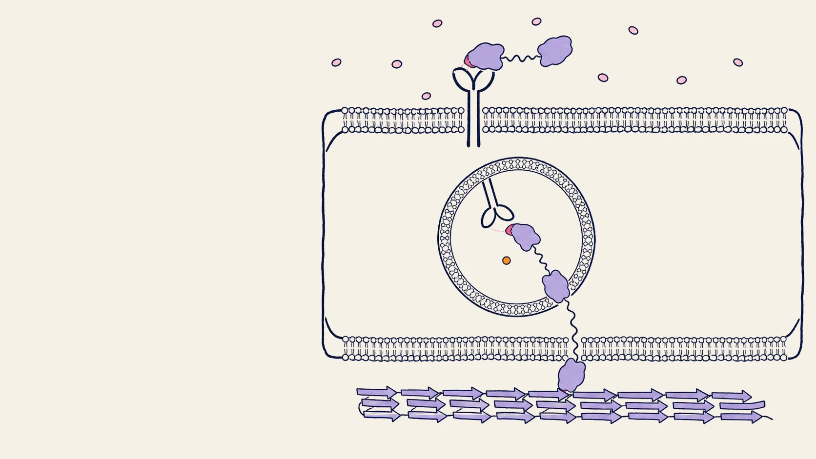
The blood-brain barrier blocks virtually all large-molecule therapeutics without active transport. Conventional antibodies are too big. Even nanobodies, among the smallest functional antibody formats, derived from camelid heavy-chain-only antibodies, don't cross freely. The established trick: hijack the transferrin receptor (TfR1), which shuttles iron across the BBB via receptor-mediated transcytosis. Bind TfR1, ride the vesicle across, release on the other side.
But "bind and release" requires pH-sensitive engineering. The nanobody must grip TfR1 at blood pH (7.4) and let go in the acidic endosome (pH 5.5). Too tight in the endosome, and the molecule gets degraded instead of transcytosing. A common engineering strategy is to introduce histidine residues at the binding interface, which protonate at low pH and weaken binding through electrostatic repulsion.
Using the Crane Protein Intelligence Platform, we designed a bispecific construct from scratch without prior nanobody sequences: one VHH domain for TfR1 transcytosis and one for alpha-synuclein fibril binding. De novo CDR design, pH-switch engineering, and a multi-stage validation pipeline produced 32 variants for the lab.
A known route: our contribution is the molecule
None of the delivery biology here is novel, and that is deliberate. Receptor-mediated transcytosis across the BBB is a validated, de-risked route: the nanobody-TfR1 complex is taken up into a vesicle on the blood side, carried across the endothelial cell, and released on the brain side. There is no pore and nothing squeezes through; it is the cell's own vesicular machinery, and the cargo's size is irrelevant next to the ~100 nm transport vesicle. Denali's Transport Vehicle, a TfR1 shuttle, is already in clinical trials.
The engineering rules are established too. What limits brain uptake is not how tightly you bind but how the receptor is sorted inside the acidifying endosome, recycled, transcytosed, or sent to the lysosome. The field converged on four principles: target a site that does not compete with transferrin, use moderate rather than maximal affinity, bind monovalently, and add a histidine pH-switch so the binder releases in the acidic endosome and rides the transcytotic route instead of being degraded. A monovalent shuttle raised brain target engagement 55-fold over its bivalent parent; an engineered pH-sensitive anti-TfR1 nanobody (three histidine mutations at the binding interface) reached more than 3.5% of injected dose per gram of brain. Our design targets, affinity window, monovalent VHH format, and pH-switch all follow directly from this body of work.
So our contribution is not the route or the pH-switch concept; it is the molecule. We applied these established design rules through the platform to generate the VHH domains entirely from scratch: de novo CDR loops, zero sequences borrowed from prior nanobodies, no camelid immunization, no grafting of published loops. The platform is the differentiator; the delivery mechanism is textbook, which is precisely what makes it a credible target for de novo design.
Two targets, two problems
The TfR1 arm targets the apical domain of human transferrin receptor 1 (PDB 1SUV). The ectodomain is a well-folded, ~670-residue globular protein, structurally cooperative, computationally tractable. The design goal: moderate affinity (Kd 100-500 nM) with a histidine-based pH-switch delivering >5-fold Kd shift between pH 7.4 and 5.5.
The alpha-synuclein arm is harder. Alpha-synuclein is intrinsically disordered as a monomer; it has no stable fold. The therapeutic target isn't the monomer at all, but the fibril: a repeating beta-sheet lattice where dozens of chains stack into a cross-beta structure. The NAC core (residues 68-79) forms an L-shaped hydrophobic motif present in every patient-derived fibril polymorph, though the surface-exposed geometry differs between polymorphs. This motif is absent in beta-synuclein, giving built-in selectivity. The design objective is fibril binding via avidity across the repeating NAC lattice.
100 nanobody sequences, 100 unique CDR3 loops, zero borrowed from nature.
100 nanobodies, designed from nothing
The platform designed 50 de novo VHH candidates per target using IgGM, a diffusion-based model that co-designs CDR sequences and structures conditioned on the antigen. Each candidate shares a common VHH framework scaffold. The CDR1, CDR2, and CDR3 loops were designed from scratch against the target structure with explicit epitope specification.
The computational funnel
Designing candidates is the easy part. The question is which ones work.
Structural validation (NbForge): All 100 candidates fold into valid nanobody structures. pLDDT above 85, NbFrame scores above 0.90. A 100% pass rate here isn't surprising: the discrimination happens downstream.
Thermostability (TEMPRO): General-purpose tools fail on nanobodies: FoldX predicts VHH Tm with r = 0.03 (essentially random). TEMPRO, a nanobody-specific predictor trained on 567 experimentally characterized VHH domains, achieves R² = 0.714 on its benchmark set. At a Tm > 65°C threshold: 41/50 aSyn and 40/50 TfR1 candidates pass.
Isoelectric point (IPC 2.0): If the nanobody's pI falls between pH 5.5 and 7.4, it crosses its own isoelectric point during endosomal transit: net charge approaches neutrality, which can increase self-association risk. Result: 40/50 aSyn and 45/50 TfR1 candidates are safe.
Aggregation (Aggrescan3D): Both lead candidates passed with strongly negative A3D scores at the exposed VH-VL interface positions, confirming IgGM correctly placed hydrophilic hallmark residues.
After thermostability and pI filtering, 34 aSyn and 36 TfR1 candidates advanced to downstream aggregation and binding analysis.
Alpha-synuclein as a monomer doesn't have a structure for the nanobody to bind. The epitope only exists in the stacked fibril lattice.
Binding validation: the fibril context problem
For TfR1, binding was scored with IntelliFold v2 and Boltz-2. The two predictors place a binder independently, so we required agreement on the actual 3D pose, not just the confidence score: each candidate's complex from both predictors was superimposed back into the full transferrin receptor assembly (the TfR1 homodimer plus iron-loaded transferrin, PDB 1SUV), and a design was kept only if it docks on the membrane-distal apical surface, clear of the receptor body, in both predicted poses. Two designs cleared this cross-predictor, full-receptor bar; both engage the accessible apical epitope, the patch that is genuinely surface-exposed on the intact receptor rather than at a buried domain interface.
For alpha-synuclein, binding validation nearly broke the pipeline. IntelliFold on a single aSyn chain gave ipTM 0.18-0.21 across all candidates, complete disorder. The fix: present Boltz-2 with a 3-chain fibril fragment. The model recognizes the homo-oligomeric assembly and presents a structured NAC surface. Result: 31/34 candidates score as STRONG_BINDERs, though this high pass rate likely reflects Boltz-2's confidence on homo-oligomeric assemblies rather than genuine discrimination between candidates. These scores are best interpreted as relative rankings within the aSyn arm.
Engineering the pH-switch
A TfR1 shuttle has to let go in the acidic endosome, not just grip in the blood. De novo binders optimized purely for affinity rarely carry the histidines that drive pH-dependent release, so the pipeline engineers them in. PROPKA3 maps the protonation landscape of a binder at both pH 7.4 and 5.5; ThermoMPNN-D scans all 138 positions for histidine tolerance, with most CDR positions tolerating histidine at predicted ΔΔG above −1.0 kcal/mol (convention: positive = stabilizing).
Cross-referencing the stability scan with the IntelliFold and Boltz-2 contact maps isolates the histidine-tolerant positions that also sit at the binding interface. Those become a focused single/double/triple mutation library, a panel of pH-switch variants that trade a small, predicted stability change for endosomal release. This step is applied to binders once they are confirmed at the bench, so the histidine budget is spent only on scaffolds that actually engage the receptor. Whether any design delivers a real Kd shift requires dual-pH SPR confirmation.
Some will fail. That's the point of 32, not 3.
32 variants, two FASTA files
The TfR1 panel contains the two designs that pass the cross-predictor, full-receptor steric check, binders that engage the accessible apical epitope of the intact receptor in both predicted poses. The aSyn library contains 30 variants: the top 30 by computational binding score, with 30 unique CDR3 sequences spanning IgGM's design diversity.
What the pipeline told us
- Fibril targets need multi-chain context. Single-chain alpha-synuclein is invisible to structure predictors. The epitope only exists in the stacked fibril lattice. This isn't a model failure; it's an input failure.
- General tools fail on nanobodies. NetSolP scores 0/100 VHH candidates above threshold. FoldX predicts Tm with r = 0.03. Domain-specific tools are required for credible results.
- Thermostability and binding are uncorrelated. The highest-Tm candidate ranked 15th by binding. Don't pick leads by Tm alone.
- Cross-tool consensus is the most reliable ranking. Candidates scoring STRONG on both IntelliFold and Boltz-2 are the highest-confidence leads.
- Validate against the whole target, and make predictors agree on the pose. Targets are often trimmed to a domain to make design tractable, but the final binder is checked against the intact assembly (dimer, ligand, and glycosylation sites) and kept only when two independent structure predictors agree on where it docks. A design that looks bound on an isolated domain but collides with the rest of the receptor, or that two predictors place differently, is caught computationally, before synthesis.
What happens next
32 variants were prioritized for gene synthesis and first-pass experimental screening. SPR will test TfR1 engagement on the full receptor, with dual-pH SPR for the pH-switch round that follows. ThT assays will test aSyn fibril engagement. DSF will validate Tm predictions. SEC-MALS will check for aggregation.
We are not developing these candidates as therapeutics. This project shows the computational workflows we deliver as services. The same capabilities are available to teams with their own targets and their own development programs.
The lab results will be the next chapter.
Translational risks
- TfR1 binding ≠ BBB delivery. Productive transcytosis depends on epitope, affinity, valency, and intracellular trafficking route, including endothelial retention versus lysosomal sorting.
- Species cross-reactivity. Human TfR1 epitopes may not cross-react with rodent or NHP orthologs.
- Bispecific formatting. Linker architecture and domain orientation are downstream steps not addressed here. Assembly may alter both arm affinities.
- Fibril polymorphism. Designed against a single fibril context. Strain coverage across polymorphs is unvalidated.
- pH-switch magnitude. The actual Kd shift may be too small for productive transcytosis.
- Developability gaps. Humanization risk, chemical liabilities, protease sensitivity, polyreactivity, and formulation behavior are not addressed.
- Immunogenicity. VHH domains are camelid-derived. Anti-drug antibodies are a risk for chronic CNS dosing.
- Target validation. Alpha-synuclein fibril clearance as disease-modifying is a hypothesis, not established biology.
Best fit
This workflow is for teams that need de novo nanobody candidates against targets where no prior binder exists, especially when the target involves structural complexity (disordered proteins, fibrillar assemblies, pH-dependent binding).
It is not a substitute for wet-lab validation. The output is a computationally ranked, mechanistically annotated variant panel. The value is in compressing the design-to-synthesis cycle and making each synthesis order count.
What we need from you: target structure or cryo-EM map, epitope hypothesis, binding constraints. What you get back: a computationally ranked variant library (typically 2-3 weeks for the computational phase) with per-candidate mechanistic annotations and explicit risk flags. Gene synthesis and wet-lab validation are separate steps.