Designing a TNF-alpha Binder That Lets Go in the Acid
1,402 generated designs, one lead lineage with a repeated chemical rationale for letting go in the acid, and twenty sequences headed to the Anthropic x Adaptyv lab. We expect to see the pH effect there.
- Evidence type
- Computational design campaign
- Published
- October 2026
13 October 2026 / computational panel / awaiting lab testing
Our protein engineers used the Crane Protein Intelligence Platform, with Linus supporting design orchestration and analysis, to prepare twenty sequences for the conditional TNF-alpha challenge: bind soluble TNF-alpha at pH 7.4, bind it less well at pH 6.0. We believe the lead lineage will show that pH effect in the lab. There are no wet-lab measurements yet, so binding, affinity and acidic release are untested until the results arrive.
- 1,402
- unique designs generated
- 62
- full-trimer complex predictions
- 4 of 4
- chemistry snapshots favor release for the lead
A binder that holds in blood and lets go in the acid
TNF-alpha is a classic drug target, and a stubborn one. The active protein is a trimer: three identical chains of 157 residues locked into a single assembly. A binder has to recognize that whole object, not one chain pulled out of context.
The task comes from the Anthropic x Adaptyv protein design challenge on Proteinbase. It added a twist. The binder should hold on at the pH of blood, 7.4, and loosen its grip at pH 6.0, roughly the acidity of an early endosome. Engineers build that behaviour into antibodies on purpose, because a molecule that releases its target in an acidic compartment can be recycled instead of consumed with it.
Nothing in a standard structure prediction knows what pH is. Folding models place atoms, they do not titrate them. So the work was really two problems in one: find a binder, then find the chemistry that lets it go.
Start wide, then check everything against the real assembly
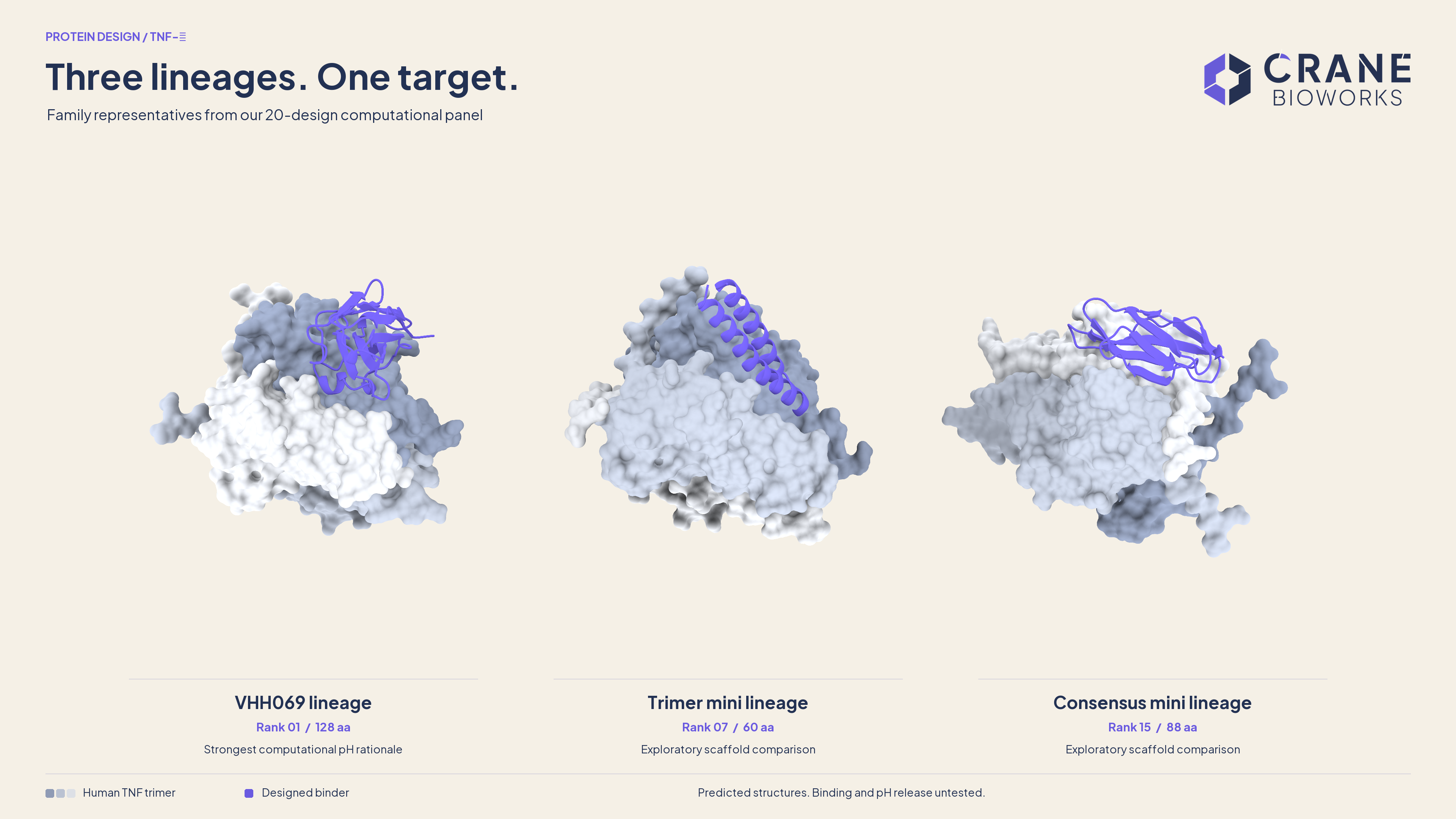
Linus generated 1,402 unique designs in two formats: nanobodies, built on a fixed framework with redesigned loops, and compact single-chain miniproteins of 60 to 95 residues. Five generation roots fed the final panel.
Every serious check ran against all three TNF chains plus the binder, using two independent structure predictors, ESMFold2 and Protenix-v2, plus mouse TNF as a second species. The reason is a lesson learned early. An earlier route that showed designs only a single TNF chain looked great: 15 of 16 sampled candidates then collided with the two chains it had not been shown. After that, the full trimer stayed in the loop.
One lineage rose to the top
The lead started as a nanobody called VHH069. It recognized TNF in a plausible pose, but its chemistry pointed the wrong way: it buried an acidic group on TNF, Glu146, and the calculations favored holding on at low pH, the opposite of the brief.
Histidine is the amino acid whose charge flips right around pH 6 to 7, which makes it the natural switch. Linus probed a pair of histidines at positions 104 and 111, then redesigned the surrounding loops in three rounds of 128 designs each, holding the framework and the histidine pair fixed. Every child was rebuilt and rechecked from scratch. Nothing inherited its parent's scores.
One child, child19, kept its recognition across both predictors and across human and mouse TNF. It became the anchor: rank 1, and the parent of most of the VHH069 family.
A histidine only helps if the rest of the interface lets it. Keeping the two histidines did not keep the pH preference.
Reading the chemistry, pose by pose
We compared the bound complex with the separated partners using PROPKA 3.5.1, which estimates how strongly each ionizable group holds a proton. In the rank 1 model, binder His104 sits at pKa 6.66 on its own but drops to 5.65 inside the complex. His111 falls from 6.43 to 4.60. At pH 6.0 the separated histidines are mostly protonated, roughly 82% for His104 and 73% for His111. Bound, they are not, roughly 31% and 4%.
Put plainly: inside the complex the histidines are held neutral. Once the partners separate they can pick up protons, and that pull grows as the solution acidifies. In this model that favors letting go. Summing every titratable group at the interface gives a factor above one when the neutral complex is favored. Across four separate structural snapshots rank 1 scores 7.41, 2.12, 2.92 and 4.37, all in the intended direction. We think this is the strongest pH-release hypothesis in the panel, and its mechanism is specific enough for the lab to confirm. It is a prediction with a mechanism behind it, not a measured release rate.
| Candidate | What the models say | How we treat it |
|---|---|---|
| Rank 1, VHH069 child19 | Repeated human and mouse support. Four of four chemistry snapshots favor release (7.41, 2.12, 2.92, 4.37). | The lead hypothesis, with coordinate assumptions stated. |
| Rank 2 | Same direction in three poses (1.43, 1.18, 4.41), but target Glu146 cancels much of the histidine gain and one repeat sits near 1. | A weaker, geometry-sensitive hypothesis. |
| Rank 10 | Both histidines help in every pose, yet opposing acidic groups flip the net result: 1.98 in the first pose, 0.370 and 0.297 in repeats. | Kept as the counterexample that shows why the net matters. |
| Ranks 9 and 11 | One supportive pose each (2.75 and 2.63), but mouse and Protenix predictions disagree sharply. | Limited exploratory probes. |
| Fourteen other designs | Every available static pH factor points the wrong way. | Scaffold and recognition comparisons, labeled as such. |
A panel built to teach us something
The final twenty are unchanged, model-generated sequences. Eleven descend from the VHH069 lineage. The rest come from independent routes: a trimer-aware miniprotein family, an 88-residue scaffold chosen because two predictors agreed on a shared alternative pose, and two further generation roots. They span 60 to 128 residues.
Most of the panel is deliberately not a bet on the lead. If the lab finds binding in an unexpected family, or finds that the histidines were the wrong lever, the evidence for that sits right next to the sequence that produced it. The score table keeps every repeat, every disagreement and every missing value visible, so family diversity is never mistaken for uniform pH evidence.
Two confidence scores, one clear signal
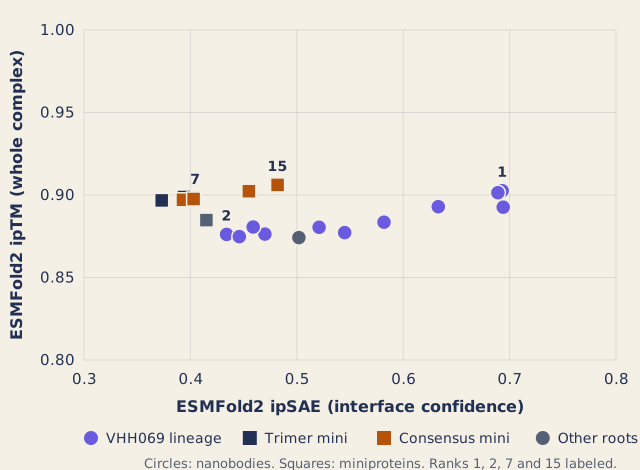
Both scores come from ESMFold2 predictions of each binder on the human TNF trimer. ipTM is scored over the whole four-chain complex, so the trimer's own contacts count too, and it sits between 0.87 and 0.91 for all twenty designs. That says the trimer folds well, not that the binder is good. ipSAE reads only the interface between binder and TNF, and it spreads the panel from 0.37 to 0.69.
Ranks 1, 9 and 12 end up almost level on ipSAE, at 0.69. Cross-model checks and the chemistry separated them: rank 1 held up across human, mouse and Protenix predictions, rank 9 collapsed on mouse and Protenix, and rank 12 had weak mouse support and no Protenix check.
Explore four of the twenty
Choose a candidate and drag the model. Slate chains are the human TNF trimer, violet is the designed binder. These are the first human ESMFold2 models recorded for each design, not selected for the best-looking pose.
Lead hypothesis: strongest combined recognition and pH rationale.
Rank 1 sequence, 128 amino acids
EVQLVESGGGLVQPGGSLRLSCAASGFPFDKYAMGWFRQAPGKGRELVAAISSDGTETYYPDSVEGRFTISRDNAKRMVYLQMNSLRAEDTAVYYCAATGSNGHNTSALRHEGSWSYWGQGTQVTVSS
What the lab will decide
Everything here is computational. Confidence scores are not affinity, the pH factors are not measured dissociation ratios, and two short target-only simulations were context rather than ranking evidence. We have no wet-lab data on any of these sequences, and we say so in the submission too.
We are optimistic anyway. The lead has a specific, testable reason to release in acid, and we expect the lab to show that pH effect. If it does not, the other families give us clear next questions instead of a dead end.
What we do have is a repeatable loop: generate widely, rebuild every candidate against the real assembly, ask a second model, read the chemistry residue by residue, and keep the disagreements. It runs on whatever target and condition you bring, whether that is a different pH, a second species, or a format constraint. The lab results will tell us which parts of the loop earned their keep.