AI Models Can Tell You Which Scaffolds Fit, But Not Which Ones Are Selective
We tested four co-folding models on two covalent inhibitors across KRAS and EGFR. The models position ligands at covalent bond distance from reactive cysteines, but treat that geometry as acceptable, not reactive. The ~1 Å distance gap is a useful diagnostic for covalent encounter geometry, not a reactivity model.
- Evidence type
- Use Case
- Published
- April 2026
Why these benchmarks
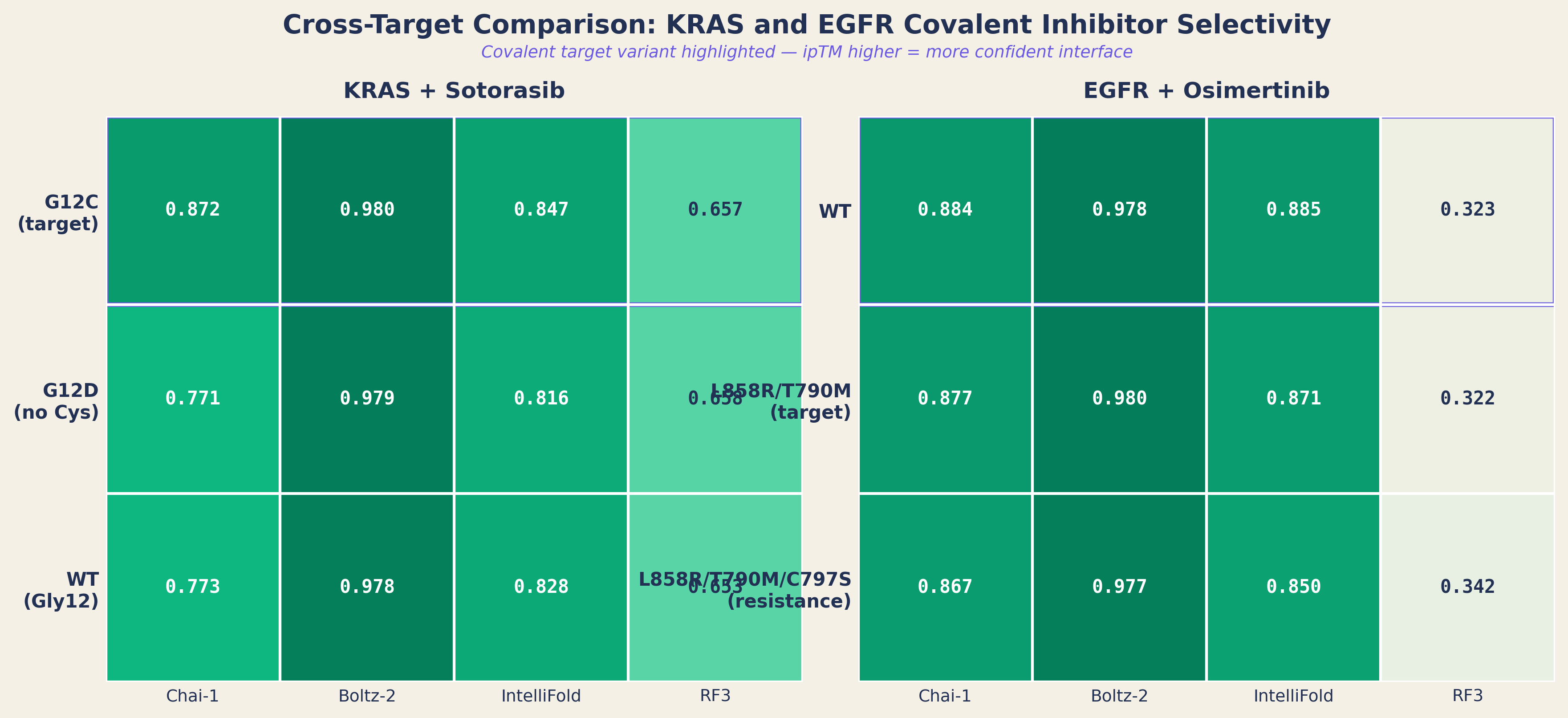
Sotorasib (Lumakras, Amgen) and osimertinib (Tagrisso, AstraZeneca) are covalent inhibitors targeting KRAS G12C and EGFR respectively. Both form irreversible thioether bonds with a cysteine, Cys12 for sotorasib, Cys797 for osimertinib. Both have well-characterized resistance mutants where the cysteine is replaced (G12D for KRAS, C797S for EGFR), eliminating the covalent mechanism while preserving the pocket.
This gives us matched decoys at two targets: variants where the scaffold should still fit the pocket, but the chemistry that makes the drug selective cannot fire. The question: can non-covalent co-folding resolve covalent selectivity?
Most of the apparent KRAS variant selectivity was memorization
All four tools easily distinguish sotorasib from a caffeine negative control (ipTM margins of 0.35-0.45), confirming basic binder vs non-binder discrimination.
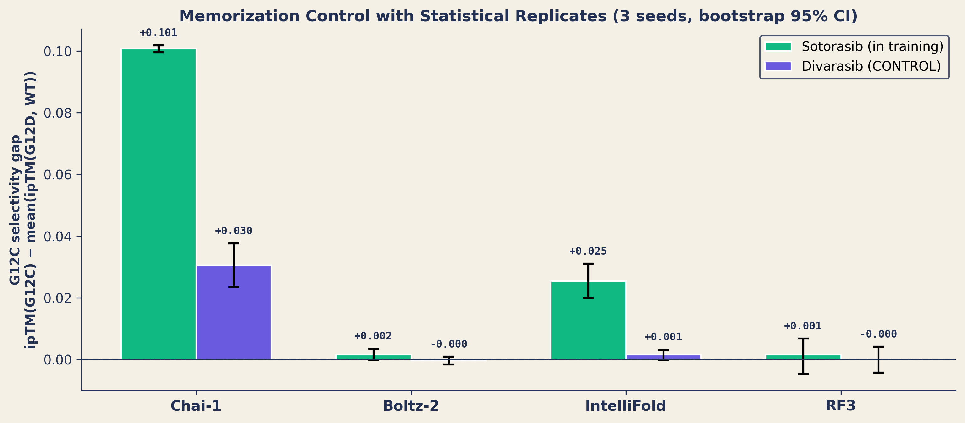
When we tested sotorasib across six KRAS variants (G12C, G12D, G12V, G12R, G13D, and wild-type), Chai-1 appeared to discriminate G12C from the other five: ipTM 0.873 vs 0.773, a 10% gap. But sotorasib's co-crystal structure (PDB 6OIM, deposited April 2019, released November 2019) predates the training cutoffs of all four models.
We repeated the experiment with divarasib, a second KRAS G12C inhibitor whose co-crystal (PDB 9PZY, released March 2026) is post-cutoff for all tools. We preregistered the primary endpoints before running the unseen control. With 3 seeds per condition and bootstrap 95% confidence intervals:
| Tool | Sotorasib gap | Divarasib gap | 95% CI (divarasib) |
|---|---|---|---|
| Chai-1 | +0.101 | +0.031 | [+0.024, +0.038] |
| IntelliFold | +0.026 | +0.002 | [-0.000, +0.003] |
| Boltz-2 | +0.002 | -0.000 | [-0.002, +0.001] |
| RF3 | +0.002 | -0.000 | [-0.004, +0.004] |
~70% of the original signal was memorization. Chai-1 retained a small, statistically significant residual: +0.031 ipTM with a 95% CI that excludes zero. The other three tools show no discrimination.
The discrimination artifact manifests as ligand misplacement: Chai-1 and IntelliFold place sotorasib 14-16 Å from the pocket for non-G12C variants: high ipTM coexisting with a completely misplaced ligand. With unseen divarasib, all tools place the ligand in-pocket for all variants. Boltz-2 consistently finds the correct pocket regardless of compound or variant, but assigns uniformly high confidence (~0.98) in this benchmark: accurate poses, unreliable confidence scores. We use it for geometry, not as a confidence filter.
The 2 Å signal: what the models actually learn
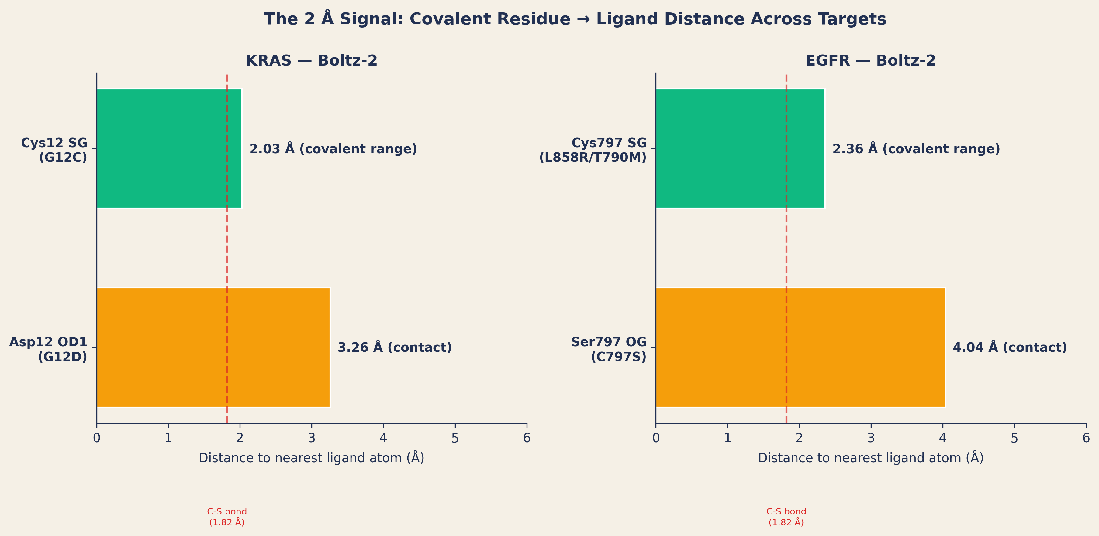
We next looked beyond confidence scores and measured the actual distance between the covalent binding residue and the nearest ligand atom. Across both KRAS and EGFR, a consistent pattern emerged:
| Target | Cys → ligand | Non-reactive → ligand | Difference |
|---|---|---|---|
| KRAS (Boltz-2) | 2.03 Å | 3.21 Å | +1.18 Å |
| KRAS (Chai-1) | 1.75 Å | 3.60 Å | +1.85 Å |
| EGFR (Boltz-2) | 2.36 Å | 4.04 Å | +1.68 Å |
| EGFR (Chai-1) | 1.70 Å | 4.40 Å | +2.70 Å |
Cysteine → ligand: 1.7-2.4 Å: consistently at covalent bond distance (C-S thioether bond = 1.82 Å). Non-reactive residue → ligand: 3.2-4.4 Å: pushed back 1-2 Å to accommodate the different side chain.
The models have learned that cysteine's thiol group gets very close to acrylamide warheads. They position the warhead at bond-formation distance for Cys and subtly reposition it for Asp or Ser. This is consistent with plausible non-covalent pre-reactive encounter geometry.
But they cannot take the next step. The co-folding models treat both 2.03 Å and 3.21 Å as acceptable geometric arrangements. The ~1 Å distance gap is a useful diagnostic for identifying pre-reactive encounter geometry, but it is not a reactivity model. Closing that gap requires covalent docking tools like CovDock (Schrödinger) or DOCKovalent, which evaluate approach angle, steric accessibility, and warhead-residue reaction geometry to distinguish "reactive contact" from "close but inert." That is the handoff point between Phase 1 and Phase 2.
PoseBusters independently confirms it
We validated all predicted poses with PoseBusters. Every pose passed 21 out of 22 chemical geometry checks. The single failure: minimum_distance_to_protein, and it fails with a perfect pattern:
| Variant type | Chemistry checks | Distance check |
|---|---|---|
| Every Cys variant (G12C, L858R/T790M) | 21/21 PASS | FAIL |
| Every non-Cys variant (G12D, C797S, WT) | 21/21 PASS | PASS |
PoseBusters independently flags the same short-contact geometry under its non-covalent plausibility checks: the Cys→warhead distance (1.7-2.4 Å) triggers a steric clash warning. This is expected: for a non-covalent complex, that distance is a clash; for a pre-reactive encounter, it is exactly where the thiol and warhead should be. A covalent-aware pipeline should treat this failure at known covalent sites as expected rather than disqualifying.
The two-phase workflow
These results (replicated across two targets, four tools, and two chemistry classes) point to a practical workflow for covalent drug design:
Phase 1: Scaffold-pocket accommodation. Use co-folding models to assess whether a molecular scaffold complements the target pocket. Verify with pocket occupancy checks and pose plausibility validation. This phase answers: does this shape belong in this pocket?
Phase 2: Covalent mechanism (chemistry). Selectivity in covalent programs comes from warhead-residue chemistry: the reactive group, the nucleophilic residue, and the geometry between them. This requires covalent docking tools (CovDock, DOCKovalent), QM calculations for warhead reactivity, or medicinal chemistry expertise. This phase answers: can we attach a warhead that reacts with the target residue and not others?
Best fit
- Scaffold triage: screen compound libraries for pocket accommodation before synthesis
- Variant panel profiling: flag pocket-shape disruptions across mutation panels
- Memorization auditing: quantify training-set retrieval vs structural prediction for your target
- Phase-gating: decide which compounds advance to covalent docking or wet-lab reactivity assays
Not a fit for: predicting clinical response, resolving covalent selectivity from co-folding alone, or replacing covalent docking where warhead-residue geometry is the primary question.
Bring a compound panel
We can run this two-phase assessment for your covalent program in 1-3 weeks: 100s of compounds, 1 target, 1 variant panel, multi-tool scoring with pocket occupancy verification and pose plausibility checks, leakage audit against your target's PDB history, and a phase-gate shortlist for covalent docking or synthesis. We ship all predicted structures (CIF/SDF), measurement scripts, and a full reproducibility report, so you can audit every result.
Also of interest: we are looking for wet-lab data to calibrate these findings, time-dependent IC50, mass-spec adduct confirmation, or kinact/KI measurements for covalent inhibitors across variant panels. If you have existing data or would be interested in running experiments alongside our computational analysis, we would like to hear from you.