Designing a Selective KRAS G12D Binder From Scratch
A de novo binder with a structurally characterized selectivity mechanism, designed entirely in silico through the Crane Protein Intelligence Platform.
- Evidence type
- Use Case
- Published
- February 2026
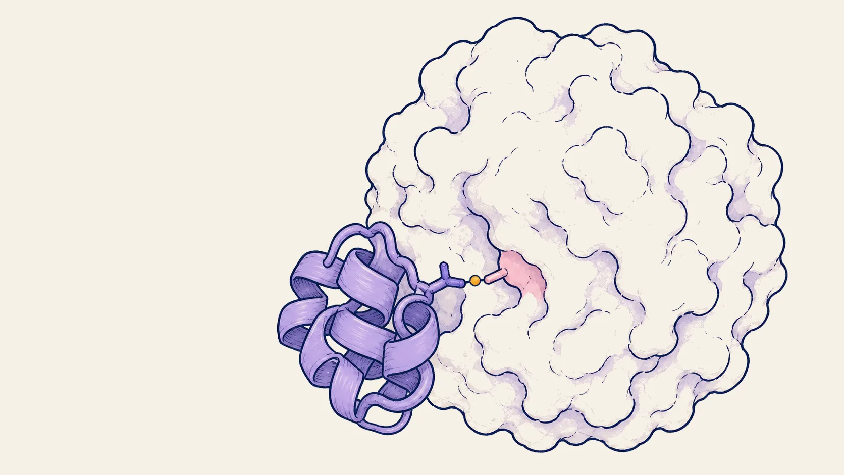
The Crane Protein Intelligence Platform orchestrates a team of specialized AI models (for structure prediction, protein design, molecular docking, and more) guided by human protein engineers. You describe your goal in plain language; the platform plans the campaign, selects the right tools, and runs the computation. This story follows one such campaign from start to finish.
The target nobody can drug
KRAS G12D is the most common cancer-driving mutation in human tumors. It fuels pancreatic, colorectal, and lung cancers, some of the deadliest malignancies worldwide. A related mutation (G12C) already has approved drugs, but G12D has none. The chemical properties of the mutated amino acid make it a poor target for traditional small-molecule drugs.
The mutation is deceptively subtle: a single amino acid swap on a nearly flat protein surface. Worse, cancer cells carrying this mutation also produce the normal (wild-type) KRAS protein. Any therapeutic must tell the two apart based on that one-residue difference, like finding a single changed letter in a 189-letter word.
This is not a problem that brute-force screening can solve. It requires understanding the 3D shape and electrical charge of the protein surface, and how the region around the mutation (called the P-loop) shifts shape over time. Exactly the kind of problem the platform was built to investigate.
The conversation
The campaign began not with a pipeline, but with a conversation. We gave the platform a one-line brief: design a protein that selectively binds KRAS G12D over wild-type KRAS. The first response asked the fundamental question: What are we trying to build, and why? It then guided a discussion across the structural biology of KRAS, the therapeutic landscape, and the biophysical constraints that would shape the design strategy.
The platform reviewed the scientific literature and distilled four design principles. In plain terms, four ideas from recent research about how to build a protein that grabs the mutant but ignores the wild-type:
- Recognize the mutation directly: Published binders achieve selectivity by making physical contact with the mutated amino acid (Asp12). One existing drug, MRTX1133, achieves 700-fold selectivity this way.
- Build a pocket that reads the target: Baker Lab's designed binders use an engineered cavity shaped to recognize specific amino acids on KRAS, like a lock and key.
- Account for protein flexibility: Proteins are not rigid. Designing against multiple shapes (conformations) rather than a single snapshot doubles the success rate (60% vs 30%).
- Use electrical charge as a filter: The mutated residue carries a negative charge that the normal glycine does not. A positively charged amino acid (arginine) on the binder can form an attractive charge-charge bond, but only with the mutant.
These principles weren't just catalogued; they informed every downstream decision. The platform translated them into measurable success criteria using ipTM, a score from 0 to 1 that predicts how confidently two proteins bind each other. The targets: high binding confidence (>0.6) with the mutant, low (<0.5) with wild-type, a clear gap between them, and 5-20 candidates worth testing in the lab.
The campaign: 340 stages, four strategies
Rather than a single linear pipeline, the platform proposed a branching campaign with four parallel strategies, each exploring a different angle of attack. The full campaign executed 340 compute stages across all strategies, running continuously across the full campaign. Zero failures.
Protein design has two layers: the backbone (the 3D shape of the protein chain) and the sequence (which amino acids sit at each position). The platform used RFdiffusion to generate backbone shapes and LigandMPNN to fill in the amino acid sequences, two specialized AI models selected from its toolkit for this task.
Round 1: Eight initial backbone shapes established baseline expectations. The best binding confidence was ipTM 0.255, well below the 0.6 target. But the data revealed something critical: the shapes that bound tightest were not the same ones that discriminated mutant from wild-type. Binding and selectivity came from different designs. This decoupling would define the rest of the campaign.
Plan A took the most selective backbone and explored different amino acid sequences on it, 16 variants. The hypothesis: the shape already encoded selectivity; the right sequence would unlock binding strength.
Plan B attacked from the other direction: 128 new backbone shapes, this time focused tightly on the mutation site. Both strategies ran in parallel.
The breakthrough: model_3_s11
Plan A produced the lead candidate: model_3_s11, sequence variant 11 on the most selective backbone shape. The platform validated it using Chai-1, a structure prediction model that co-folds two proteins to assess whether they bind. The result: binding confidence of 0.522 against the mutant, but only 0.314 against wild-type, a clear selectivity gap. Of the 16 sequence variants on the same backbone, the other 15 scored far lower. A single standout, confirming that aggressive sequence sampling on the right backbone shape pays off.
Across the full campaign, 135 candidates were scored. 78 showed some selectivity for the mutant. Four exceeded the overall quality threshold. model_3_s11 was the only one that met all criteria: strong binding and clear selectivity. The branching strategy, exploring both backbone shapes and amino acid sequences in parallel, was essential for finding a candidate that balances both.
Anatomy of a selective binder
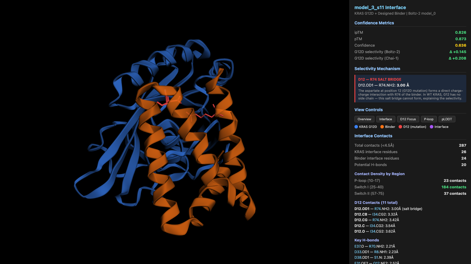
To verify the result, the platform submitted model_3_s11 to a second, independent structure prediction model: Boltz-2. Using a different algorithm reduces the risk that one model's biases produced a false positive. Boltz-2 agreed: binding confidence of 0.826 against the mutant, 0.681 against wild-type: same direction, even stronger signal. Two independent methods, same conclusion.
287 contacts, one critical bond
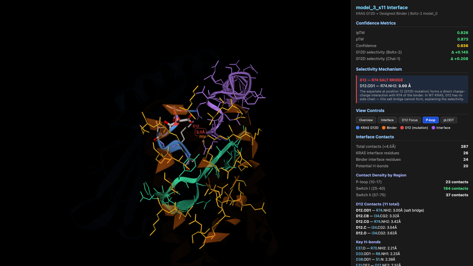
The designed binder is 80 amino acids long and makes 287 physical contacts with KRAS G12D, including 20 hydrogen bonds. It wraps around the mutation site (the P-loop) and two neighboring regions, gripping the target with a broad, stable interface.
The selectivity mechanism: D12-R74
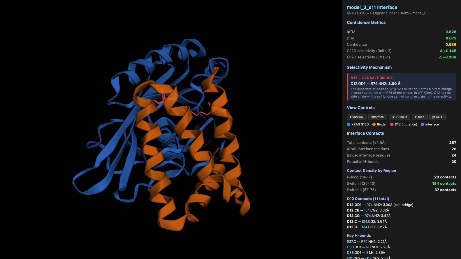
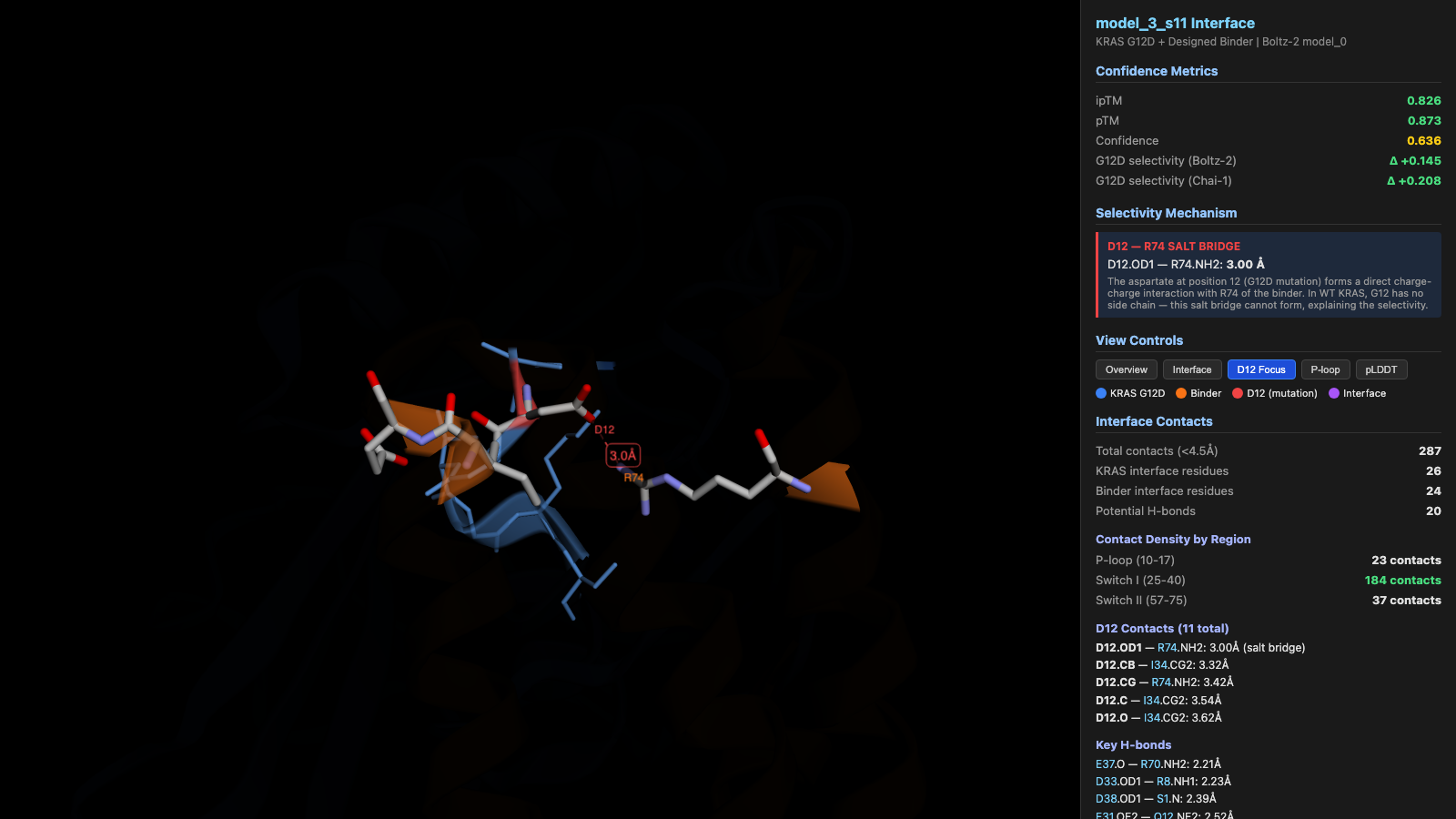
Here is the core finding, and why selectivity works. The mutated amino acid (aspartate at position 12) carries a negative electrical charge. The binder places a positively charged amino acid (arginine at position 74) directly opposite it, at a distance of just 3.00 Å (less than a billionth of a meter). Opposite charges attract: this forms a strong bond called a salt bridge.
In normal (wild-type) KRAS, position 12 is glycine, the smallest amino acid, with no side chain at all. There is physically nothing for the binder's arginine to grab. The salt bridge can only form with the mutant. This is the structural explanation for selectivity, and it emerged directly from the design principles the platform identified at the start.
The binder makes 5 direct contacts with the mutation site, all within 3-4.3 Å. Not a single tenuous interaction: a focused contact cluster, exactly as predicted.
The dissent: IntelliFold v2
Science demands skepticism. Two models agreed, but agreement could reflect shared biases. The platform selected a third, independent structure prediction model, IntelliFold v2, specifically to challenge the result. (The Crane Protein Intelligence Platform orchestrates 30+ specialized AI models and selects the right ones for each task. For this validation step, it chose three models with different architectures to maximize the chance of catching false positives.)
IntelliFold v2 disagreed: it predicted low binding confidence for both mutant and wild-type, with no selectivity signal.
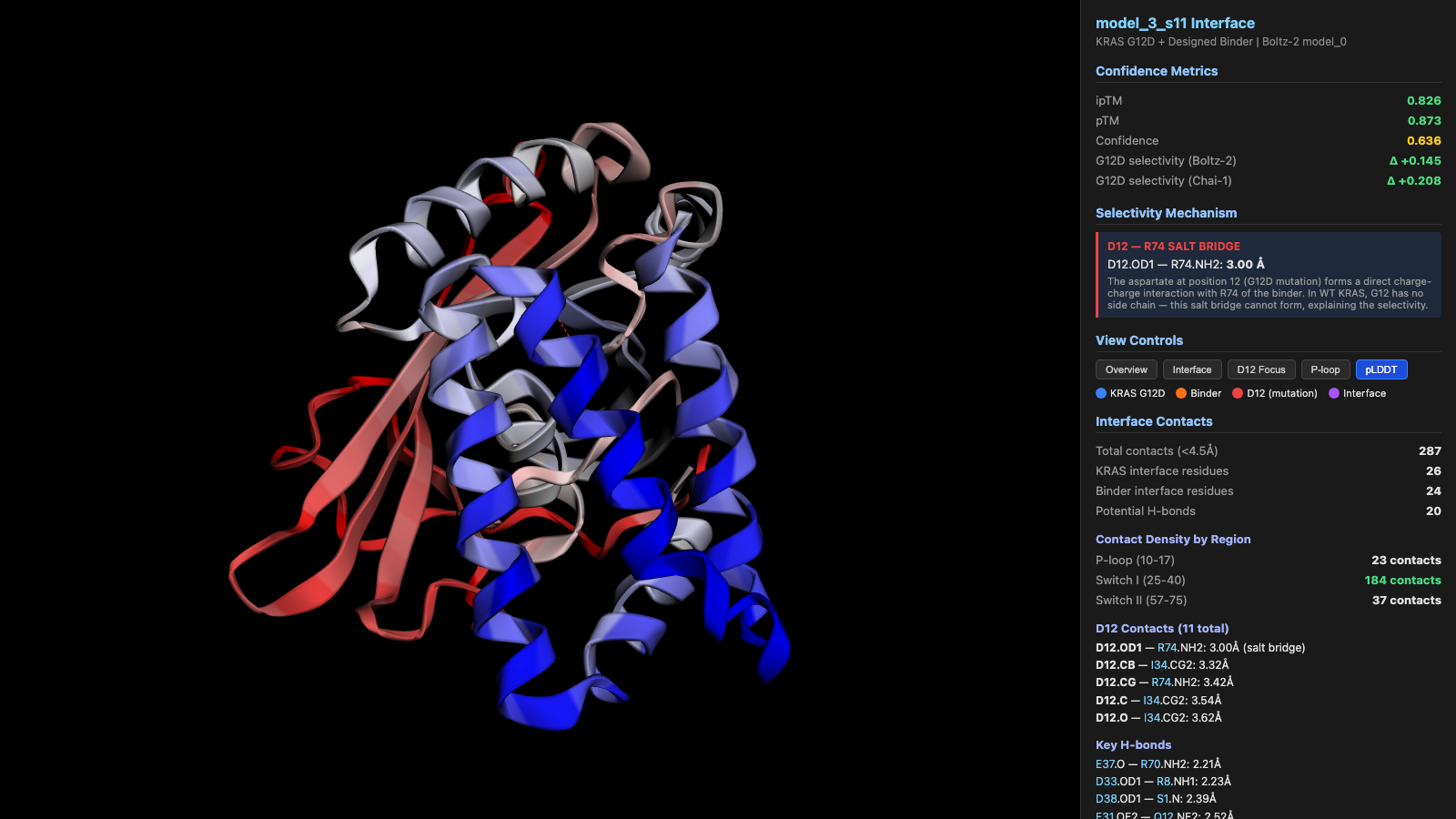
This disagreement is scientifically important. IntelliFold v2's low confidence in the binder's structural stability (pLDDT 0.52, a score measuring how reliably each part of the protein folds) suggests uncertainty about whether this 80-amino-acid designed protein holds its shape. The KRAS target itself folds reliably across all three models. The honest interpretation: model_3_s11 shows a promising binding mode with a clear selectivity mechanism confirmed by two methods, but binder stability is an open question, a well-characterized risk for designed-from-scratch proteins that informs the next round of optimization.
The numbers
Pipeline stages executed
340
Stage failures
0
Candidates scored
135 (G12D + WT pairs)
Unique backbones explored
136+
Structure prediction methods
3 (Chai-1, Boltz-2, IntelliFold v2)
Interface contacts (lead)
287 (including 20 H-bonds)
Selectivity mechanism
D12-R74 salt bridge, 3.00 Å
What this shows
This campaign ran entirely on a computer through the Crane Protein Intelligence Platform. No wet lab. No physical experiments. The output: a protein binder candidate with a structural explanation for how and why it selectively binds the cancer-driving mutant, validated by multiple independent prediction methods.
Traditional computational protein design is sequential: define target, generate shapes, design sequences, score, filter, repeat. In this campaign, the decision-making happened through continuous dialogue. When the first round revealed that binding strength and selectivity came from different designs, the platform proposed two parallel strategies. When Plan A's lead candidate emerged, the platform immediately ran independent validation while Plan B was still running. When a third model disagreed, it documented the dissent as a scientific finding rather than dismissing it.
Every observation documented. Every result scored against success metrics defined at the start. 340 stages, zero failures, one conversation.
What comes next
model_3_s11 is not a drug. It is a computationally validated hypothesis, with a clear structural rationale, reproducible predictions from two of three methods, and a known risk (stability) that can be addressed in the next iteration: optimizing the binder to fold more reliably, advancing more of the selective candidates through validation, and running molecular dynamics simulations to confirm the key salt bridge holds up over time.
From problem statement to mechanism-explained lead candidate, one conversation, zero failures. This is what AI-driven protein engineering looks like when the right platform is in place.
Try it with your target
The Crane Protein Intelligence Platform can run a design campaign like this for your protein, in days, not months. Describe your target, get computational candidates with structural rationale, test the top hits in your lab, and feed the results back for the next cycle. Each round sharper than the last.